Health
How can I tell if my dog has an eye problem?
Just with his particularly expressive eyes, your dog conveys his love, excitement, sadness, apprehension, or a whole range of other emotions.
Share the article:
However, if your dog has eye problems, they may no longer be able to express their emotions as clearly. Some conditions can quickly lead to lasting damage, ranging from changes in the structure of the eye to complete loss of vision. It is important to recognise eye conditions in your dog as quickly as possible, because if your vet intervenes early, they can often help achieve the best possible recovery.
What is the structure of a dog’s eye?
To identify an eye problem in a dog, it is important to understand the specific structure of their eye.
Your dog’s eye, like that of most mammals, is shaped like a globe. The cornea is the outermost layer of the eye and helps protect it. When it is not damaged, this layer is completely transparent and allows you to see your dog’s pupil in the center of the coloured iris.
Your dog’s eyeball is divided into two fluid-filled chambers separated by the lens. The lens is essential for vision because it focuses light rays from the outside onto the retina, which lines the back of the eye.
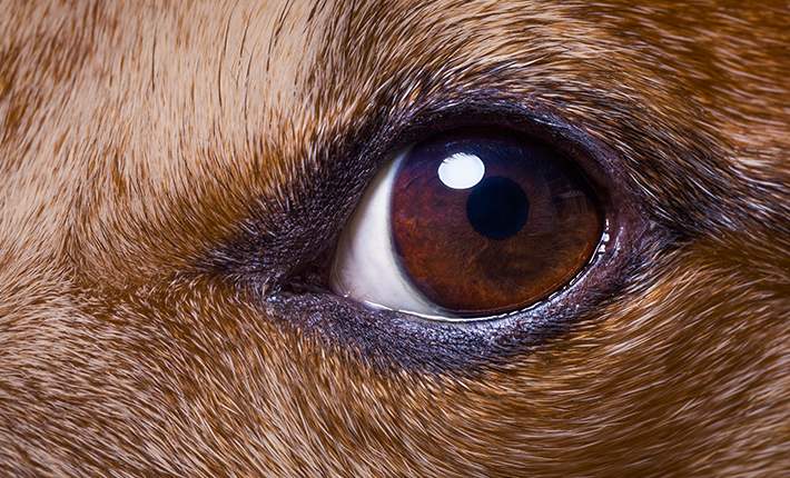
Unlike humans, dogs have three eyelids: a lower eyelid, an upper eyelid, and a well-developed third eyelid called the “nictitating membrane.” Located in the lower part of the inner corner of the eye, between the lower eyelid and the eyeball, it plays an important protective role because it can cover the eye. It also has a mechanical function and produces part of the tear film, helping to keep the eye hydrated and lubricated.
Normally, the nictitating membrane is not visible because it is hidden under the lower eyelid, but it becomes visible when there is a problem. This is known as “protrusion of the nictitating membrane.” It is therefore important to be familiar with your dog’s eyes, as some changes are not always obvious.
The different types of eye conditions in dogs
Several eye conditions of varying severity can present with similar clinical signs, and only your vet will be able to make the correct differential diagnosis. On your own, do not attempt any self-medication, apart from cleaning around the eyes with saline solution, before getting veterinary advice.
- If the surface of your dog’s eye takes on a whitish appearance, there is probably swelling of the cornea. This may be caused by an injury from a foreign body, an ulcer, glaucoma, or a viral infection.
- If your dog has a half-closed, watery eye, with the nictitating membrane, the pink membrane that starts from the inner corner of the eye, partially covering the eyeball, this may indicate the early stages of glaucoma, but also an ulcer or a foreign body such as a grass seed.
- If both eyes are watery, with swollen, red eyelids, an infection or allergy is probably present.
As a general rule, an early diagnosis and rapid intervention are often essential to save the eye and prevent serious lasting damage.
Some conditions can indeed cause serious lasting damage if they are not treated in time. For example, acute glaucoma can cause your pet to lose vision in only 48 hours. Foreign bodies that go unnoticed under the third eyelid, or ulcers, can damage the cornea and lead to vision loss.
The prognosis and functional recovery after treatment of the eye and its associated structures often depend on how quickly treatment begins after symptoms appear. This is why any eye condition, even one that seems mild, should be shown to your vet without delay.
Here are some common eye conditions in dogs:
Conjunctivitis in dogs
Inflammation of the conjunctiva is a very common condition in dogs and can have many causes, such as allergies, bacterial or viral infections, or irritation. In cases of conjunctivitis, the eye is red and the tissues surrounding the cornea are swollen, sometimes slightly oedematous. The eyelids may also be swollen and painful. The dog may blink frequently and tend to rub their face. There may also be discharge.
Because conjunctivitis can have many causes, some of which are infectious, you should not wait to consult your vet. They will perform the necessary examinations to identify the cause of the inflammation and prescribe the appropriate treatment, for example:
- by removing the source of irritation, such as a misplaced eyelash or foreign body, and administering eye drops to relieve local pain
- by prescribing antibiotics and eye rinses in the event of infection
- or by prescribing an anti-allergy treatment
Epiphora: my dog has a watery eye
This is an excessive flow of tears, meaning your dog seems to be crying all the time. The clear tears that run from the eye can eventually stain the wet fur brown or rust-colored or give it an unpleasant smell.
Epiphora develops when there is:
- excessive tear production, caused for example by poorly positioned eyelashes, conjunctivitis, or a corneal ulcer;
- poor tear drainage, for example when the tear ducts are blocked.
Very small dog breeds, such as miniature or toy breeds, appear to be predisposed, often because of poor tear drainage.
Your vet will carry out the necessary examinations to identify the cause of the epiphora and recommend an appropriate treatment.
What is entropion in dogs?
Some dogs may have eyelids that roll inward, a condition called entropion. Their eyelashes then irritate the cornea, causing excessive tear production, while the inward rolling blocks the tear drainage system, or tear ducts, preventing tears from draining normally. This problem is often hereditary and can be corrected surgically.
Dry eye: my dog has dry eyes
Here, it is the opposite: the eye is dry because not enough tears are being produced.
Glaucoma in dogs: a threat to their eyesight
Glaucoma develops when there is an imbalance between the normal production of aqueous humor, the fluid that fills the chamber in front of the lens, and insufficient drainage of that fluid. The amount of fluid increases, raising the intraocular pressure, or IOP.
Glaucoma is accompanied by numerous clinical signs that you can easily identify:
- A dilated pupil, noting that the two pupils may not be the same size if glaucoma affects only one eye
- One eye, or both eyes, may also appear larger and more protruding because of the increase in volume
- Redness of the eye or dilation of the blood vessels in the white part of the eye
- Pain, sometimes severe, may cause your dog to keep their eyes partially closed or squinted, or to rub them
- Lethargy, tiredness, or even loss of appetite
- Loss of vision
If your dog shows any signs of glaucoma, you must consult your vet immediately. This is an absolute eye emergency, and prompt care is needed to prevent vision loss.
Glaucoma is diagnosed by measuring intraocular pressure, or IOP, using a device called a tonometer.
Your vet will then recommend the appropriate treatments to reduce the IOP and keep it within normal physiological values, either through the use of eye drops or through surgery.
Cataracts in dogs
Just like humans, dogs can develop cataracts. A cataract is an opacity of the lens that prevents light from passing through properly. As a result, light does not reach the retina and its receptors as effectively, leading to vision loss.
Cataracts are often hereditary, and some breeds are predisposed, such as the Australian Shepherd, French Bulldog, Staffordshire Bull Terrier, and others.
This condition can appear with age, as dogs get older, just like people, but it can also develop in cases of diabetes, malnutrition, radiation exposure, inflammation, or trauma.
How can I tell if my dog has a cataract?
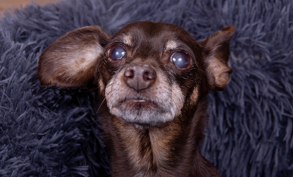
A lens affected by a cataract appears whitish or bluish. This opacity, which can be easily seen by looking at your dog’s pupil, may however be confused with lens sclerosis, a normal thickening and hardening of the lens with age. Only a vet can distinguish between the two conditions.
How does a cataract progress in dogs?
It may remain stable or worsen until it completely prevents vision. Whether a cataract progresses or stabilises depends on several factors, such as the type of cataract, the breed, or the presence of other risk factors. If vision loss is gradual, the dog will generally adapt by relying much more on their other senses, especially hearing and smell.
Many dogs adapt to their cataract without treatment. However, cataract surgery, performed by vet specializing in ophthalmology, can allow them to regain good vision. Speak with your vet, who can refer you to a specialist if your dog needs one.
Luxation, or prolapse, of the tear gland of the nictitating membrane, known as the “nictitating gland,” in dogs
The nictitating membrane contains a gland that can become visible when inflamed; this is known as luxation of the nictitating gland. This condition is characterized by the appearance of a swollen red or pink mass, ranging in size from a small pea to a small cherry, at the inner corner of the eye. The luxation may be intermittent, meaning it appears and disappears, or permanent.
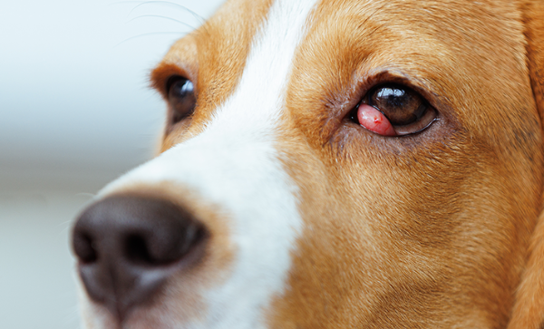
As soon as you notice the first symptoms, contact your vet so treatment can be started. Intermittent luxation may be relieved with anti-inflammatory eye drops. If it is permanent, surgical treatment under general anesthesia is required.
How can I relieve my dog’s eye discomfort?
Eye conditions share many common clinical signs, so it is not always easy for an owner to tell what is urgent and what is not. For example, conjunctivitis and glaucoma do not have the same visual prognosis and are not treated in the same way.
That is why, if you have the slightest doubt about an eye problem in your dog, you should consult your vet promptly.
References
Looking for a vet?
Your vet plays a big role in your pet’s health. Enter your location and get a list of vets near you.
In the spotlight
Wondering if your cat has fleas? Here are 9 telltale signs
What are the symptoms of fleas in cats, and how can you recognize them? Discover the 9 common signs of fleas in cats and learn how to get rid of them!
9 signs your cat has fleas
How can you tell if your cat has fleas? We’ll reveal the main symptoms so you can take the right steps to get rid of them.
Vaccination schedule for dogs & cats
Wondering what the cat vaccination schedule looks like? What are the most essential dog vaccines that your dog should get? Learn more here!
Canine ear infection: What is it and how is it treated?
Can you recognize the symptoms of ear infection in your dog? Find out by reading this article.
Fleas and ticks: is my pet at risk?
Your cat needs prompt attention if these parasites make an appearance.
Gastritis in cats
Symptoms, Causes, and Treatment of Gastritis in Cats
Everything you need to know about spaying your female dog
Sterilization of female dogs is one of the most widespread procedures for controlling unwanted litters and preventing certain pathologies such as mammary tumors and uterine infections.
The low-down on leptospirosis
One of the risks that have been increasingly reported in many countries worldwide that affects dogs is leptospirosis, also known as “lepto.”
Mange in cats: Everything you need to know
What is mange in cats, how is it transmitted, what is its treatment and how can you prevent it?